Meningomyelocele (MMC), also known as Myelomeningocele, is the most severe and most common form of spina bifida. It is a congenital birth defect in which the spinal cord, nerves, and protective membranes (meninges) protrude through an opening in the bones of the spine, forming a fluid-filled sac on the baby’s back.
Because the spinal nerves are exposed or damaged, children with meningomyelocele may experience weakness or paralysis of the legs, bladder and bowel dysfunction, orthopedic deformities, and hydrocephalus (excess fluid in the brain).
MMC develops during the first month of pregnancy when the neural tube fails to close completely. Early diagnosis, prompt surgical repair, and long-term multidisciplinary care are essential to reduce complications and improve the child’s quality of life.
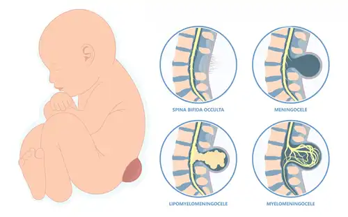
The symptoms depend on the size and location of the spinal defect.
Common symptoms of meningomyelocele in children include:
The higher the spinal defect, the greater the likelihood of neurological impairment.
Meningomyelocele develops during early fetal growth when the neural tube does not close properly.
Common causes and contributing factors include:
Most cases result from a combination of genetic and environmental factors.
Meningomyelocele is classified according to the location of the spinal defect.
Certain factors increase the risk of meningomyelocele.
Risk factors include:
Adequate folic acid supplementation before conception and during early pregnancy significantly reduces the risk.
Without proper treatment, meningomyelocele can lead to lifelong complications.
Many children develop excess cerebrospinal fluid in the brain, which may require a VP shunt or Endoscopic Third Ventriculostomy (ETV).
Part of the brain extends downward into the spinal canal, causing swallowing, breathing, or neurological problems.
Damage to the spinal nerves may affect movement of the legs.
Children may develop urinary incontinence, urinary retention, or recurrent urinary tract infections.
Constipation and loss of bowel control are common.
Children may develop clubfoot, scoliosis, hip dislocation, and joint deformities.
Reduced sensation increases the risk of pressure sores and skin ulcers.
Some children may experience cognitive or developmental challenges, particularly if hydrocephalus is present.
Many cases are diagnosed before birth through routine prenatal screening.
Diagnosis may include:
These tests help determine the severity of the defect and identify associated conditions such as hydrocephalus and Chiari II malformation.
Treatment focuses on protecting the spinal cord, preventing infection, and managing associated complications.
Most babies undergo surgery within the first 24–48 hours after birth.
During surgery:
Early surgery reduces the risk of infection and protects the exposed nerves.
Children who develop hydrocephalus may require:
Some children may require treatment for:
Treatment may include:
Many children benefit from:
A multidisciplinary approach provides the best long-term outcomes.
Recovery depends on the level of the spinal defect and associated neurological problems.
Parents are usually advised to:
Long-term follow-up is essential to monitor growth and neurological development.
Helpful care measures include:
Consistent rehabilitation and supportive care help children achieve maximum independence.
Parents should seek immediate medical attention if the child develops:
Early medical evaluation can prevent serious complications.
Meningomyelocele is the most severe and most common form of spina bifida, where the spinal cord and nerves protrude through an opening in the spine.
Yes. Most cases are diagnosed during pregnancy through ultrasound and maternal AFP screening. Fetal MRI may be used in selected cases.
Yes. Early surgical repair is recommended to protect the spinal cord, reduce infection risk, and prevent further nerve damage.
Walking ability depends on the level of the spinal defect. Some children walk independently, while others require braces, walkers, or wheelchairs.
Hydrocephalus commonly occurs because meningomyelocele is often associated with Chiari II malformation, which interferes with the normal flow of cerebrospinal fluid.
Taking 400–800 micrograms of folic acid daily before conception and during early pregnancy significantly reduces the risk of neural tube defects, including meningomyelocele.
Dr. Muni Varma is an experienced Pediatric Neurosurgeon and Pediatric Surgeon specializing in the diagnosis and treatment of meningomyelocele (MMC) and other complex congenital neurological disorders in children. With expertise in neonatal spinal surgery, hydrocephalus management, VP shunt procedures, minimally invasive pediatric surgery, and multidisciplinary rehabilitation, Dr. Varma provides comprehensive child-focused care using advanced diagnostic and surgical techniques. From prenatal counseling and early surgical repair to long-term bladder, bowel, orthopedic, and neurological management, he focuses on preserving nerve function, preventing complications, and helping children achieve their best possible quality of life. Early expert intervention and continuous follow-up provide the foundation for healthy growth, improved mobility, and long-term well-being.