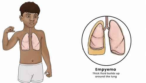
Empyema is a condition in which pus collects in the pleural space, the thin space between the lungs and the inner wall of the chest. In children, empyema most commonly develops as a complication of bacterial pneumonia, when infection spreads from the lung into the pleural space.
The accumulated pus can compress the lung and make breathing difficult. Common bacteria associated with pediatric empyema include Streptococcus pneumoniae, Staphylococcus aureus, and Group A Streptococcus.
Empyema requires prompt medical evaluation. Treatment generally involves antibiotics, supportive care, and drainage of infected pleural fluid when the infection is complicated or the child is significantly unwell.
Symptoms often develop during or after pneumonia and may continue despite appropriate antibiotic treatment.
Common symptoms of empyema in children include:
A child with pneumonia who continues to have fever or breathing difficulty after around 48 hours of appropriate antibiotic treatment may need evaluation for a pleural effusion or empyema.
Empyema usually develops when an infection in the lungs spreads into the pleural space.
Common causes include:
Pneumonia remains the most common underlying cause of empyema in children.
Pleural infection can progress through different stages.
Fluid collects around the lung during pneumonia but is not infected. A small uncomplicated effusion may improve with appropriate antibiotic treatment.
The infection begins to involve the pleural fluid. The fluid may become infected and develop septations or loculations.
Pus or infected fluid accumulates in the pleural space. Drainage is often required along with antibiotics.
The infected fluid becomes divided into pockets by fibrin and scar tissue. This can make drainage more difficult and may require additional procedures or surgery.
Certain conditions can increase the risk of developing a complicated pleural infection.
Risk factors include:
However, empyema can occur in an otherwise healthy child following pneumonia.
Without appropriate treatment, empyema can cause serious complications.
Pus and fluid around the lung can compress it and interfere with normal breathing.
Reduced lung expansion may cause hypoxaemia.
The infection can spread into the bloodstream and cause a severe systemic infection.
A collection of pus may develop within the lung.
The infected fluid can form separate pockets, making drainage more difficult.
Long-standing inflammation can cause thickening and scarring of the pleural lining, restricting lung expansion.
Complicated empyema may require hospitalization, drainage procedures, and a longer course of antibiotics.
Diagnosis is based on the child’s symptoms, physical examination, and imaging studies.
Tests may include:
Chest ultrasound is particularly useful for determining whether a pleural effusion is simple, complicated, or loculated. CT is not routinely required but may be considered when the child does not improve or another underlying condition is suspected.
Treatment depends on the child’s clinical condition and the stage of the pleural infection.
Antibiotics are an essential part of treatment because empyema is usually caused by bacterial infection. The choice of antibiotic depends on the suspected organisms, local resistance patterns, and results of cultures or other tests.
A child may require:
Children with significant respiratory distress or complicated effusions generally require hospital treatment.
If infected fluid or pus has accumulated, a chest tube or other drainage catheter may be inserted to remove the infected material and allow the lung to expand.
In selected children with loculated pleural infection, medicines that help break down fibrin and improve drainage may be used through the chest tube.
VATS is a minimally invasive surgical procedure that can be used when drainage and medical treatment are insufficient or when there is significant loculated infection.
During VATS, the surgeon can:
The exact approach is decided according to the child’s clinical condition and response to initial treatment.
Most children recover well with timely treatment, although recovery may take several weeks depending on the severity of the infection.
Parents are usually advised to:
Children who have required drainage or surgery may need additional follow-up to ensure that the lung has fully recovered.
Helpful care measures include:
Do not stop antibiotics early even if the child appears to be feeling better.
Parents should seek urgent medical attention if the child develops:
Severe breathing difficulty, low oxygen levels, or signs of sepsis require emergency medical care.
Empyema is an accumulation of infected fluid or pus in the pleural space surrounding the lung, most commonly occurring as a complication of bacterial pneumonia.
No. Pneumonia is an infection of the lung tissue, while empyema is an infected collection in the pleural space around the lung. Empyema can develop as a complication of pneumonia.
Yes. Empyema commonly develops when bacterial pneumonia spreads into the pleural space.
No. Treatment depends on the stage and severity. Some children respond to antibiotics and drainage, while others with complicated or loculated empyema may require VATS or another surgical procedure.
Recovery varies depending on the severity of infection and treatment required. Children with uncomplicated disease may recover relatively quickly, while complicated empyema may require a longer hospital stay and follow-up.
Most children recover well with appropriate treatment. However, severe or prolonged infection can cause pleural scarring and restricted lung expansion, making early treatment important.
Recurrence is uncommon after successful treatment, but persistent or recurrent symptoms require medical reassessment to ensure that the infection has completely resolved.
Dr. Muni Varma is an experienced Pediatric Surgeon specializing in the diagnosis and treatment of complex chest and respiratory surgical conditions in children. With expertise in pediatric thoracic surgery, minimally invasive procedures, chest drainage, and management of complicated infections, Dr. Varma provides comprehensive child-focused care in coordination with pediatricians, pulmonologists, intensivists, and other specialists when required. From early assessment and stabilization to drainage procedures, VATS when indicated, and post-treatment follow-up, the approach focuses on controlling infection, helping the lung re-expand, reducing complications, and supporting a safe recovery.