An Arteriovenous Malformation (AVM) is an abnormal connection between arteries and veins in which blood flows directly from the arteries into the veins without passing through the normal network of tiny blood vessels called capillaries.
Normally, arteries carry oxygen-rich blood to tissues through smaller blood vessels, and veins then carry the blood back to the heart. In an AVM, this normal pathway is bypassed. As a result, blood can flow through the abnormal vessels at high pressure, which may damage nearby tissues or cause bleeding.
AVMs can develop in different parts of the body, including the brain, spinal cord, head and neck, chest, abdomen, or limbs. The symptoms and treatment depend largely on where the AVM is located, its size, and whether it has caused bleeding or other complications.
Some AVMs are present from birth but may not become noticeable until later in childhood. Early evaluation is important because certain AVMs can enlarge or cause serious complications over time.
The symptoms depend on the location and size of the AVM.
Common symptoms may include:
Brain or spinal AVMs may remain unnoticed until they cause bleeding, seizures, headaches, or neurological symptoms.
The exact cause of most AVMs is not known.
AVMs are believed to develop because of abnormal formation of blood vessels during fetal development. In some cases, genetic changes affecting blood vessel development may contribute to their formation.
Possible contributing factors include:
An AVM is generally not caused by something a child or parent did during pregnancy.
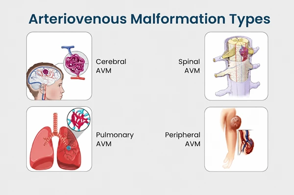
AVMs can be classified according to the part of the body they affect.
A brain AVM is an abnormal connection between arteries and veins within or around the brain. It may cause headaches, seizures, neurological problems, or bleeding inside the brain.
A spinal AVM develops in or around the spinal cord and can affect movement, sensation, or bladder and bowel function.
These AVMs can affect the face, scalp, mouth, or neck and may cause visible swelling, skin discoloration, bleeding, or enlargement of the affected area.
An AVM may develop in an arm or leg and cause swelling, pain, skin changes, or abnormal growth of the affected limb.
AVMs can occasionally develop in organs such as the lungs, liver, or gastrointestinal tract and may cause bleeding or other organ-specific symptoms.
Most children with AVM do not have identifiable risk factors.
Factors that may be associated with AVMs include:
In most cases, an AVM occurs sporadically without a known family history.
Some AVMs remain stable, while others may cause significant complications.
Fragile abnormal blood vessels can rupture and cause bleeding. Brain AVM bleeding can result in a hemorrhagic stroke.
Brain AVMs can irritate surrounding brain tissue and cause seizures.
Bleeding or pressure from an AVM may cause weakness, numbness, difficulty speaking, vision problems, or problems with coordination.
AVMs in the limbs or other parts of the body may cause persistent pain and swelling.
Large superficial AVMs may cause skin discoloration, ulcers, or bleeding.
Very large AVMs can allow a substantial amount of blood to bypass normal circulation and, in rare severe cases, place extra strain on the heart.
Diagnosis depends on the location of the suspected AVM.
Doctors may use:
Angiography provides detailed information about the arteries, abnormal connections, and draining veins and can be particularly important when planning treatment.
Treatment depends on the AVM’s location, size, blood-flow pattern, symptoms, and risk of complications. Some AVMs can be monitored, while others require active treatment.
Small or low-risk AVMs that are not causing symptoms may sometimes be monitored with regular clinical examinations and imaging.
Embolization is a minimally invasive procedure in which a catheter is guided through the blood vessels to the AVM. Special materials are then used to reduce or block abnormal blood flow.
Embolization may be used:
Surgery may be recommended when the AVM can be safely removed.
The surgeon removes the abnormal blood vessels while preserving nearby healthy tissues and important blood vessels.
For selected brain AVMs, focused radiation can be used to gradually close the abnormal blood vessels. The effect occurs over time rather than immediately.
Some complex AVMs require more than one treatment method. For example, embolization may be performed first to reduce blood flow, followed by surgical removal or another definitive treatment.
The treatment plan should be individualized by a multidisciplinary vascular or neurosurgical team.
Recovery depends on the location of the AVM and the treatment performed.
After treatment, children may require:
Children treated for brain or spinal AVMs may need additional monitoring for neurological symptoms.
Home care depends on the child’s treatment and the location of the AVM.
Helpful measures include:
Parents should not ignore new neurological symptoms or unexplained bleeding.
Immediate medical attention is required if a child develops:
These symptoms may indicate bleeding or another serious complication.
An AVM is an abnormal connection between arteries and veins that bypasses the normal capillary network.
Many AVMs develop because of abnormal blood vessel formation during fetal development, although symptoms may not appear until childhood or later.
Most established AVMs do not simply disappear on their own. Some may remain stable, while others can enlarge or become symptomatic.
No. Treatment depends on the location, size, symptoms, and risk associated with the AVM. Some can be monitored, while others require embolization, surgery, radiosurgery, or a combination of treatments.
Yes. Some AVMs can rupture and cause significant bleeding. The risk depends on the location and characteristics of the AVM.
No. An AVM is a high-flow abnormal connection between arteries and veins, while a hemangioma is a different type of vascular lesion with a different natural history and treatment approach.
Many children do well after appropriate treatment, particularly when the AVM is diagnosed before serious complications occur. Long-term follow-up may be necessary depending on the type and location of the AVM.
Dr. Muni Varma is an experienced Pediatric Surgeon specializing in the evaluation and management of complex congenital and vascular conditions in children. For children with arteriovenous malformations, he focuses on accurate diagnosis, careful assessment of the AVM’s location and extent, and coordinated treatment planning with the appropriate vascular, interventional, or neurosurgical specialists. Depending on the condition, treatment may involve observation, minimally invasive procedures, surgical management, or a combination approach. With a child-focused approach and expertise in complex pediatric surgical conditions, Dr. Varma aims to reduce complications, preserve normal function, and provide appropriate long-term follow-up.