Pediatric Occipital Encephalocele Repair Surgery is a specialized procedure used to repair an opening in the skull at the back of the head and manage the sac containing cerebrospinal fluid, protective brain coverings, and, in some children, neural tissue.
The main goals of surgery are to protect the brain and neural structures, manage the encephalocele safely, close the skull and dural defect, prevent cerebrospinal fluid (CSF) leakage, and achieve secure soft-tissue coverage.
The surgical technique depends on the size of the encephalocele, the contents of the sac, the size of the skull defect, the relationship with the brain and venous structures, and the child’s neurological condition.
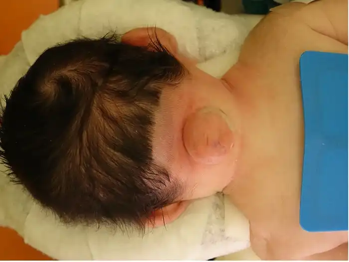
Surgery may be recommended when an occipital encephalocele is present and requires structural repair.
The surgical team evaluates:
The timing and surgical plan are individualized according to the child’s condition.
The main objectives of surgery are to:
Surgery repairs the structural defect, but the child’s long-term neurological outcome depends on the amount and function of the brain tissue involved.
Before surgery, the child undergoes detailed neurological evaluation and brain imaging. MRI and other imaging studies may help the surgical team understand the contents of the encephalocele and its relationship with the brain, skull, and blood vessels.
During surgery:
The exact procedure varies significantly depending on the anatomy of the individual encephalocele.
The encephalocele sac is carefully examined during surgery.
It may contain CSF, meninges, brain tissue, or a combination of these structures.
The surgeon determines which tissue can be safely preserved and how the sac should be managed while protecting functioning brain tissue and important blood vessels.
One of the most important considerations during surgery is distinguishing functioning neural tissue from abnormal or non-functional tissue.
If functioning brain tissue is present within the encephalocele, the surgical team aims to preserve it whenever safely possible.
The exact management depends on the child’s imaging findings and surgical anatomy.
After management of the encephalocele, the protective covering around the brain, called the dura, is carefully repaired.
A secure dural closure helps:
A watertight closure is an important surgical objective.
The occipital encephalocele is associated with an opening in the skull.
Depending on the size and anatomy of the defect, the surgeon may reconstruct the skull using appropriate tissue or surgical materials.
The goal is to provide adequate protection for the underlying brain and create a stable cranial contour.
The exact reconstruction technique depends on the child’s age, skull defect, and surgical findings.
After repair of the deeper structures, the surrounding soft tissue and skin are closed carefully.
Large encephaloceles may require careful soft-tissue reconstruction to achieve adequate coverage without excessive tension.
Secure closure helps reduce the risk of wound breakdown and CSF leakage.
Some children with encephalocele also have hydrocephalus, which may require separate evaluation and treatment.
The child may undergo brain imaging and neurological assessment to determine whether excess CSF is accumulating.
If CSF diversion is required, treatment may include a ventriculoperitoneal (VP) shunt or another appropriate procedure.
The timing of hydrocephalus treatment is individualized according to the child’s neurological and imaging findings.
Occipital encephalocele repair is a complex pediatric neurosurgical procedure.
The operation can be performed safely with detailed preoperative imaging and careful surgical planning, but the risks depend greatly on the size and contents of the encephalocele and its relationship with the brain and major blood vessels.
Important factors include:
Potential benefits include:
The long-term neurological outcome depends largely on the underlying brain development and the extent of neural involvement.
Potential complications include:
The actual risks vary according to the child’s individual anatomy and neurological condition.
After surgery, the child is monitored closely.
The medical team may assess:
Children undergoing complex repair may require intensive monitoring during the early recovery period.
The length of hospital stay depends on the child’s condition and surgical complexity.
Parents should:
Urgent medical evaluation is important if the child develops persistent vomiting, seizures, unusual sleepiness, fever, wound opening, fluid leakage, or new neurological symptoms.
Long-term follow-up is important because some children may have associated neurological or developmental conditions.
Follow-up may include:
The follow-up plan is individualized according to the child’s neurological condition.
The developmental outcome after encephalocele repair varies significantly.
Some children may have relatively preserved neurological function, while others may have developmental, motor, seizure, or other neurological challenges related to the underlying brain involvement.
Regular developmental assessment can help identify areas where additional support may be useful.
The timing depends on wound healing, neurological status, the extent of surgery, and associated conditions.
Light activities can be gradually increased according to medical advice.
Activities that could cause significant impact to the head or put pressure on the surgical area may need to be restricted until adequate healing has occurred.
The treating neurosurgical team will provide individualized activity recommendations.
Some children require long-term multidisciplinary follow-up.
This may include:
The child’s needs may change as they grow, making ongoing assessment important.
The cost varies depending on the complexity of the encephalocele and the child’s medical requirements.
Factors that may affect the total cost include:
Parents should discuss the expected treatment plan and estimated expenses with the treating hospital.
Parents should discuss:
Understanding the individual anatomy helps parents prepare for the surgical procedure and recovery.
It is a specialized neurosurgical procedure used to repair an opening in the occipital skull and safely manage the encephalocele sac and any neural tissue contained within it.
An encephalocele may contain cerebrospinal fluid, protective brain coverings, brain tissue, or a combination of these structures. The contents vary between children.
No. Functioning neural tissue should be preserved whenever possible. The surgical plan is based on imaging and the relationship between the tissue, brain, and blood vessels.
Yes. Depending on the size and anatomy of the defect, the surgeon may reconstruct the skull as part of the procedure.
Some children with encephalocele also develop hydrocephalus. If CSF diversion is required, a VP shunt or another appropriate treatment may be considered.
Not every child requires a shunt. The need depends on whether hydrocephalus is present and how the child’s CSF circulation behaves.
Development varies significantly depending on the amount of brain involvement and associated neurological abnormalities. Some children may develop well, while others may require ongoing developmental and rehabilitation support.
Recovery varies according to the size and complexity of the encephalocele, the surgical reconstruction, neurological status, and associated conditions such as hydrocephalus.
Yes. Follow-up may include monitoring neurological development, head growth, hydrocephalus, seizures, and other associated conditions.
Dr. Muni Varma offers advanced Pediatric Occipital Encephalocele Repair Surgery for Children in Lucknow, India. With more than 10 years of experience, he uses a careful and child-focused approach to provide accurate evaluation, appropriate surgical planning, and better long-term care. He offers personalized treatment and follow-up support for children requiring encephalocele repair, helping parents understand the treatment process and providing quality pediatric surgical care in Lucknow, India.