A hemangioma is a benign (non-cancerous) growth made up of extra blood vessels. It is one of the most common vascular tumors seen in infants. Hemangiomas may not be visible at birth or may appear as a small red or pale mark that grows during the first few months of life.
Infantile hemangiomas commonly occur on the skin of the head, face, neck, or trunk, but they can also develop internally, including in organs such as the liver. Most hemangiomas eventually become smaller and lighter over time without causing major problems.
However, some hemangiomas can grow rapidly or develop near important structures such as the eyes, nose, mouth, or airway. These lesions may interfere with vision, breathing, feeding, or normal development and may require treatment.
Early evaluation helps determine whether a hemangioma can simply be monitored or needs medical or surgical management.
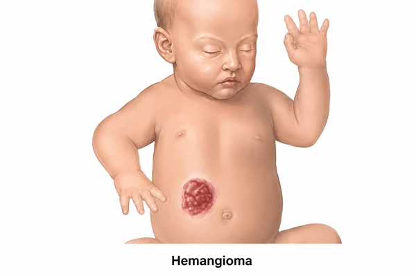
The appearance and symptoms of a hemangioma depend on its size, depth, location, and growth pattern.
Common symptoms of hemangioma in children include:
Deep hemangiomas may appear as a soft lump under otherwise normal-looking skin.
The exact cause of infantile hemangiomas is not completely understood.
Hemangiomas develop because of abnormal growth and multiplication of blood vessel cells. They are not caused by an injury or something parents did during pregnancy.
Factors associated with a higher likelihood of infantile hemangioma include:
Most children with hemangiomas do not have a family history of the condition.
Hemangiomas can be classified according to their depth, appearance, and location.
These occur close to the surface of the skin and typically appear bright red. They are sometimes described as “strawberry hemangiomas.”
These develop deeper beneath the skin and may appear bluish, purple, or skin-colored. They often feel soft and raised.
These contain both superficial and deep components and may appear as a combination of red and bluish swelling.
A single, well-defined lesion develops in one particular area.
The lesion covers a larger area and follows a particular region or developmental pattern of the skin. Segmental hemangiomas may have a higher risk of complications and associated abnormalities.
Several factors are associated with an increased likelihood of infantile hemangioma.
Risk factors include:
A hemangioma can also develop in a child without any identifiable risk factor.
Most hemangiomas are harmless, but some can cause complications depending on their location and size.
The surface of a hemangioma can break down and form an ulcer, which may be painful and susceptible to infection.
Ulcerated hemangiomas may bleed, particularly when located in areas exposed to friction.
A hemangioma near the eye may interfere with vision development or cause pressure on the eye.
Hemangiomas affecting the airway can cause noisy breathing, difficulty breathing, or airway obstruction.
Lesions around the lips, mouth, or throat can interfere with feeding.
Large or prominent hemangiomas may leave residual skin changes, such as loose skin, scarring, or discoloration after they shrink.
Children with numerous skin hemangiomas may occasionally have hemangiomas involving internal organs, particularly the liver, and may require further evaluation.
Most hemangiomas can be diagnosed by examining the lesion and reviewing the child’s medical history.
Diagnosis may include:
Imaging is particularly useful for deep, large, complicated, or unusual lesions.
Most hemangiomas do not require active treatment because they naturally enter a growth phase followed by gradual involution. Treatment is considered when a hemangioma threatens vital functions, causes significant complications, or has a high risk of permanent disfigurement.
Small, uncomplicated hemangiomas may simply be monitored.
Regular follow-up may include:
Propranolol, a beta-blocker, is the first-line systemic treatment for many problematic infantile hemangiomas.
It can help:
Treatment with propranolol should be prescribed and monitored by an appropriately trained doctor.
For selected small and superficial hemangiomas, topical medicines such as timolol may be considered.
Corticosteroids were commonly used before propranolol became standard treatment. They may still have a role in selected situations when other treatments are unsuitable.
Laser therapy may be considered for certain superficial lesions, particularly residual redness or specific ulcerated hemangiomas.
Surgical removal is not usually the first treatment for infantile hemangioma. It may be considered for:
The treatment approach should be individualized according to the child’s age, lesion characteristics, location, and risk of complications.
Recovery depends on the type of treatment and the location of the hemangioma.
Children receiving medical treatment generally require regular follow-up to assess:
After surgery, wound care and follow-up are important.
Most children with uncomplicated hemangiomas do well and gradually see the lesion become smaller and lighter as they grow.
Helpful care measures include:
Parents should not apply home remedies or unprescribed medicines directly to the lesion.
Parents should have a hemangioma evaluated if it:
A hemangioma affecting vision, breathing, or feeding requires prompt medical evaluation.
A hemangioma is a benign growth made up of extra blood vessels. Infantile hemangiomas commonly appear during the first weeks or months of life.
Many infantile hemangiomas gradually shrink and become lighter as the child grows. However, some may leave behind loose skin, discoloration, or other residual changes.
No. Most uncomplicated hemangiomas can be safely monitored. Treatment is recommended when the lesion causes or has a significant risk of causing functional problems, ulceration, or permanent disfigurement.
Propranolol is commonly used as the first-line systemic treatment for problematic infantile hemangiomas. It should only be used under appropriate medical supervision.
Infantile hemangiomas are benign and are not cancerous.
True recurrence after adequate treatment is uncommon, although some lesions may continue to change during the natural growth and involution phases.
Surgery may be considered for selected lesions that cause functional problems, leave significant residual tissue, or are unlikely to improve adequately with observation or medical treatment.
Dr. Muni Varma is an experienced Pediatric Surgeon specializing in the evaluation and management of hemangiomas and other congenital and vascular conditions in children. He focuses on assessing the size, location, depth, growth pattern, and potential complications of each hemangioma before recommending an appropriate treatment plan. Depending on the child’s condition, management may include observation, medical treatment, minimally invasive procedures, or surgery when required. With expertise in pediatric surgery and multidisciplinary care, Dr. Varma aims to protect important functions such as vision, breathing, and feeding while achieving the best possible medical and cosmetic outcome.