Retroperitoneal Teratoma is a rare tumor that develops in the retroperitoneum, the space located behind the abdominal cavity where organs such as the kidneys, pancreas, and major blood vessels are situated. It is a type of germ cell tumor that arises from primitive cells capable of developing into different types of body tissues, such as hair, skin, muscle, bone, fat, or teeth.
Most retroperitoneal teratomas in children are benign (non-cancerous), especially in infants and young children. However, some may contain immature or malignant (cancerous) cells, making early diagnosis and complete surgical removal essential.
Retroperitoneal teratomas are uncommon but are among the important abdominal tumors seen in pediatric surgical practice. With timely surgery, most children have an excellent long-term prognosis.
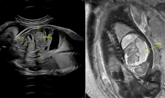
The symptoms depend on the size and location of the tumor.
Common symptoms of retroperitoneal teratoma in children include:
Small tumors may not cause any symptoms and are sometimes discovered during imaging performed for another reason.
Retroperitoneal teratomas develop before birth due to abnormal growth of primitive germ cells.
Common causes and contributing factors include:
The exact cause is not fully understood, and most cases occur without any known risk factors.
Retroperitoneal teratomas are classified based on the maturity of the tumor cells.
1. Mature Teratoma: This is the most common type in children. It contains well-developed tissues such as skin, fat, hair, bone, or teeth and is usually benign.
2. Immature Teratoma: This type contains immature or embryonic tissues and has a greater potential to behave aggressively. Additional treatment and close follow-up may be required.
3. Malignant Teratoma: A rare type in which cancerous cells are present within the tumor. These tumors often require surgery along with chemotherapy depending on the stage and pathology.
Although the exact cause remains unknown, certain factors may be associated with retroperitoneal teratoma.
Risk factors include:
Most children diagnosed with retroperitoneal teratoma do not have identifiable risk factors.
If left untreated, retroperitoneal teratoma may cause several complications.
A large tumor may press on the kidneys, intestines, bladder, or blood vessels.
Pressure on the intestines may lead to vomiting, constipation, or bowel obstruction.
Compression of the urinary tract may cause difficulty passing urine or kidney swelling (hydronephrosis).
Although uncommon, parts of the tumor may become infected.
Rarely, the tumor may rupture, causing abdominal pain and inflammation.
In rare cases, a mature teratoma may contain or develop malignant components.
Doctors use imaging studies and laboratory tests to diagnose retroperitoneal teratoma and determine its extent.
Diagnosis may include:
Imaging helps determine the size, location, and relationship of the tumor to nearby organs.
Treatment depends on the size, location, and pathology of the tumor.
Surgery is the primary treatment for retroperitoneal teratoma.
During surgery:
Complete removal provides the best chance of cure.
Children with malignant or certain immature teratomas may require chemotherapy after surgery based on the pathology report.
Regular follow-up with clinical examination, imaging, and tumor marker tests is important to detect recurrence.
Most children recover well after complete surgical removal.
Parents are usually advised to:
Recovery depends on the size of the tumor and whether additional treatment is required.
Helpful care measures include:
Regular follow-up helps ensure early detection of recurrence if it occurs.
Parents should seek medical attention if the child develops:
Early medical evaluation helps identify complications and ensures timely treatment.
A retroperitoneal teratoma is a rare germ cell tumor that develops behind the abdominal cavity and may contain tissues such as hair, fat, bone, or teeth.
Most retroperitoneal teratomas in children are benign, but some may be immature or malignant and require additional treatment.
Diagnosis usually involves ultrasound, CT scan or MRI, blood tests for tumor markers, and confirmation through histopathological examination after surgical removal.
Yes. Complete surgical removal is the standard treatment and offers the best chance of cure.
Recurrence is uncommon after complete removal of a benign tumor but may occur in immature or malignant teratomas, making regular follow-up important.
Yes. Most children with benign retroperitoneal teratomas recover completely after surgery and lead healthy, active lives.
Dr. Muni Varma is an experienced Pediatric Surgeon specializing in the diagnosis and surgical treatment of retroperitoneal teratomas and other complex pediatric abdominal tumors. With expertise in neonatal surgery, pediatric oncology surgery, minimally invasive procedures, and advanced abdominal tumor excision, Dr. Varma provides comprehensive child-focused care using modern diagnostic and surgical techniques. From accurate diagnosis and complete tumor removal to long-term follow-up and coordinated multidisciplinary care, he focuses on achieving complete tumor clearance while preserving the function of surrounding organs. Early expert treatment and regular monitoring help ensure excellent long-term outcomes and healthy growth for children.