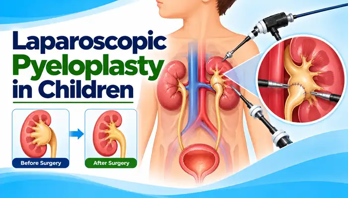
Laparoscopic pyeloplasty is a minimally invasive surgical procedure used to correct a blockage at the junction where the renal pelvis (the part of the kidney that collects urine) connects to the ureter (the tube that carries urine to the bladder). This area is known as the ureteropelvic junction (UPJ).
In children, this blockage is often caused by a congenital narrowing of the ureteropelvic junction, a condition called ureteropelvic junction obstruction (UPJO). It can prevent urine from draining normally from the kidney and may lead to swelling of the kidney, known as hydronephrosis.
During laparoscopic pyeloplasty, a pediatric surgeon makes small incisions and uses a camera and specialized instruments to remove or reshape the narrowed segment and reconnect the healthy tissues. The goal is to improve urine drainage and protect kidney function.
Laparoscopic pyeloplasty may be recommended when investigations show that a blockage at the ureteropelvic junction is significantly affecting urine drainage or putting kidney function at risk.
A child may need evaluation for this procedure if they have:
Not every child with hydronephrosis needs surgery. Mild cases may be monitored with regular examinations and imaging. The decision depends on symptoms, the severity of obstruction, kidney function, and changes over time.
The main purpose of laparoscopic pyeloplasty is to restore the normal flow of urine from the kidney into the ureter.
The procedure may help to:
The extent of improvement depends on the severity of the obstruction and the kidney’s condition before surgery. An already damaged kidney may not regain all its previous function.
Laparoscopic pyeloplasty is performed under general anesthesia. The exact surgical plan depends on the child’s anatomy, age, kidney function, and the nature of the obstruction.
The procedure generally involves the following steps:
The procedure is commonly based on the Anderson-Hynes dismembered pyeloplasty technique, although the surgeon may adapt the approach to the child’s individual anatomy.
The appropriate approach depends on the child’s condition, the anatomy of the obstruction, and the treating team’s experience.
The procedure is performed through small incisions using a camera and specialized instruments. It allows the surgeon to reconstruct the ureteropelvic junction without a large open incision.
Robotic-assisted surgery uses a robotic surgical system to control specialized instruments through small incisions. Availability depends on the hospital, equipment, and the child’s clinical requirements.
Open pyeloplasty uses a larger incision to access and reconstruct the obstructed junction. It remains an effective option in selected cases, including when minimally invasive surgery is unsuitable or unavailable.
The surgeon recommends the most appropriate technique after evaluating the child’s condition.
Before surgery, the pediatric surgical and urology team may recommend investigations to understand the obstruction and assess kidney function.
These may include:
Not every child needs every investigation. The treating team selects tests according to the child’s history, examination, and previous results.
When surgery is indicated, laparoscopic pyeloplasty offers several potential benefits:
The outcome depends on the child’s anatomy, kidney function, and the presence of other urinary tract abnormalities. Surgery cannot guarantee complete recovery of previously lost kidney function.
Laparoscopic pyeloplasty is an established procedure for appropriately selected children when performed by an experienced surgical team. However, as with any operation, it carries risks.
Possible complications include:
The treating team explains the benefits and risks based on the child’s age, medical history, kidney function, and individual surgical plan.
Recovery varies from child to child. The medical team monitors pain, hydration, urine output, and general well-being after surgery.
During recovery, parents may notice:
The length of the hospital stay depends on the child’s progress and the hospital’s protocol. Children should return to school and physical activities according to the surgeon’s instructions.
Seek prompt medical attention if the child develops fever, worsening abdominal or flank pain, persistent vomiting, difficulty passing urine, or other concerning symptoms.
Following discharge, parents should follow the care plan provided by the surgical team.
Important aftercare measures include:
A temporary stent must not be left in place longer than advised. The team will explain whether removal requires a separate procedure.
Follow-up is important because symptom improvement alone does not always confirm that urine drainage has normalized.
The doctor may recommend:
Hydronephrosis may take time to improve after a successful operation. The treating team interprets imaging findings alongside symptoms and, when needed, functional tests.
The cost of laparoscopic pyeloplasty in children in Lucknow varies according to the hospital, the child’s age and medical condition, the surgical approach, anesthesia requirements, length of hospital stay, investigations, and any additional treatment required.
Parents should request an individualized estimate from the treating hospital. The estimate should clarify whether it includes the surgeon’s fees, anesthesia, investigations, hospital stay, medicines, stent-related care, and follow-up.
Before consenting to laparoscopic pyeloplasty, parents should understand:
Parents should discuss any concerns about anesthesia, previous infections, medicines, or other medical conditions with the treating team.
Laparoscopic pyeloplasty is a minimally invasive operation that corrects a blockage where the kidney’s urine-collecting area joins the ureter. It aims to restore urine drainage and help protect kidney function.
Children may need pyeloplasty when ureteropelvic junction obstruction causes significant kidney swelling, impaired drainage, recurrent symptoms, or a risk to kidney function. Mild cases may only require monitoring.
Laparoscopic surgery uses smaller incisions and may offer advantages in postoperative discomfort, recovery, and scarring for suitable patients. Open surgery remains effective, and the best approach depends on the child’s condition and the surgical team’s expertise.
Recovery varies depending on the child’s age, surgical approach, and overall health. The child may resume routine activities gradually, following the surgeon’s instructions. Full recovery and imaging improvement may take longer than the initial return to normal activities.
A temporary ureteral stent is often used, but not every surgical plan is identical. If a stent is placed, the doctor will explain its care and the planned removal date.
Correcting an obstruction may help preserve kidney function and can sometimes improve drainage-related functional measurements. The degree of improvement depends on how much function was affected before surgery.
Persistent or recurrent obstruction is possible, although many children have satisfactory outcomes. Follow-up appointments and imaging help identify children who need further assessment or treatment.
Ultrasound is commonly used to identify kidney swelling. A diuretic renal scan may help assess urine drainage and relative kidney function. Additional investigations are selected according to the child’s clinical findings.
No. Some children have mild hydronephrosis that can be monitored without surgery. Pyeloplasty is considered when the clinical findings suggest significant obstruction, symptoms, or a risk to kidney function.
Dr. Muni Varma offers Laparoscopic Pyeloplasty in Children in Lucknow, India. With more than 10 years of experience, he uses a careful and child-focused approach to provide appropriate evaluation, surgical planning, and follow-up care. He helps parents understand why pyeloplasty may be needed, what the procedure involves, and how surgery can improve urine drainage and help protect kidney function in children with ureteropelvic junction obstruction.