Pediatric arteriovenous malformation (AVM) surgery is a specialized treatment approach used to manage abnormal connections between arteries and veins in children. Normally, blood flows from arteries through tiny blood vessels called capillaries before entering the veins. In an AVM, arteries connect directly to veins, bypassing the normal capillary network.
These abnormal connections can occur in different parts of the body, including the brain, spinal cord, skin, muscles, and internal organs. Depending on their location and size, AVMs may cause swelling, pain, bleeding, skin changes, neurological symptoms, or other complications. Some AVMs remain unnoticed until they are discovered during medical investigations.
Treatment may involve surgical removal, endovascular embolization, stereotactic radiosurgery for selected brain AVMs, or a combination of approaches. The choice depends on the AVM’s location, blood flow, size, symptoms, and risk of complications. Not every AVM requires immediate surgery, and some may be managed with careful monitoring.
Dr. Muni Varma offers pediatric surgical consultation in Lucknow for children requiring evaluation of vascular abnormalities and discussion of appropriate treatment options. Complex AVMs, particularly those involving the brain or spinal cord, require assessment by the relevant specialist team.
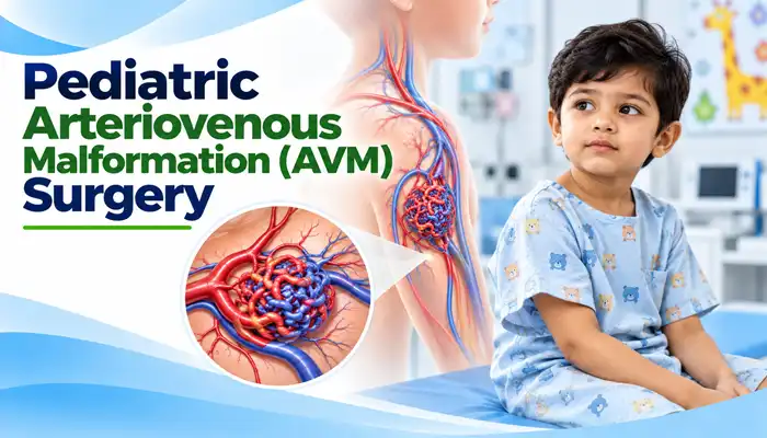
Surgery or another form of active treatment may be considered when an AVM causes symptoms, presents a significant risk of bleeding, or affects the function of nearby tissues or organs.
A child may require specialist evaluation if they experience:
The presence of an AVM does not automatically mean surgery is necessary. The treating team evaluates the risks of intervention against the risks of observation and determines the most appropriate course of action.
The primary goal of AVM treatment is to reduce the risks associated with abnormal blood flow and, when appropriate, eliminate the abnormal vascular connections.
Depending on the type and location of the AVM, treatment may aim to:
The expected benefits differ between AVMs. Treatment planning must account for the possibility that intervention itself can damage nearby blood vessels, nerves, brain tissue, or other important structures.
The procedure depends on where the AVM is located and which treatment approach has been selected.
For a surgically accessible AVM, the general process may include:
Brain AVM surgery is particularly specialized and may involve a craniotomy, microsurgical techniques, and intraoperative angiography. AVMs located in other parts of the body require different approaches based on their anatomy.
Microsurgery involves removing the abnormal vascular connections through an operation. For selected brain AVMs, the surgeon accesses the lesion through an opening in the skull and carefully separates it from surrounding brain tissue.
Microsurgical removal may provide immediate elimination of an AVM when complete removal is achieved and confirmed. However, it may not be appropriate for lesions located deep within the brain or near critical structures.
Endovascular embolization is a minimally invasive procedure used to reduce or block blood flow through an AVM. A specialist guides a thin catheter through the blood vessels to the targeted area and delivers an embolic material.
Embolization may be used before surgery to reduce blood flow and potentially make removal safer. It may also be used as part of a treatment plan for selected AVMs. In some cases, it can be a definitive treatment, but in others it provides only partial or temporary control.
Stereotactic radiosurgery uses highly focused radiation to damage the abnormal vessels within selected AVMs, particularly certain brain AVMs that are difficult to reach surgically.
Despite the term “surgery,” this technique does not involve a conventional surgical incision. The vessels close gradually over time, and the AVM may take months or years to become completely obliterated. The risk of bleeding can persist until treatment is confirmed to have succeeded.
Some complex AVMs require more than one treatment method. For example, embolization may be performed before microsurgical removal, or radiosurgery may be considered for an AVM that is unsuitable for direct surgery.
Staged treatment allows the team to plan intervention according to the lesion’s characteristics and the child’s clinical needs.
Some AVMs may be monitored rather than treated immediately when the risks of intervention outweigh the expected benefits. This decision requires specialist assessment, a clear follow-up plan, and appropriate imaging when indicated.
The choice between observation and intervention is individualized and should not be based solely on the child’s age or the apparent size of the lesion.
AVM treatment requires detailed assessment because the anatomy and blood-flow pattern can vary significantly between children.
The evaluation may include:
For complex cases, the treatment plan may involve pediatric surgery, neurosurgery, interventional neuroradiology, vascular specialists, and other relevant disciplines.
AVM surgery is a complex procedure, and its risks depend on the location, size, and anatomy of the malformation, as well as the treatment method selected.
Potential complications include:
Brain and spinal AVMs require particularly careful risk assessment because intervention can affect important neurological structures. The treating specialists should explain the expected benefits, possible complications, and alternative approaches before the family makes a decision.
When treatment is appropriate and successful, potential benefits may include:
Outcomes depend on the AVM’s anatomy, the treatment selected, and whether complete elimination is achieved. No treatment can guarantee a particular outcome for every child.
Recovery depends on the type of AVM, the location of the lesion, and the procedure performed.
After microsurgery, the child may require hospital monitoring and a period of recovery before returning to routine activities. Recovery after embolization or radiosurgery follows a different course, and the duration of observation depends on the procedure and the child’s condition.
General recovery guidance may include:
Children undergoing brain AVM treatment may need additional rehabilitation if the AVM or its treatment affects movement, speech, balance, or other functions.
Follow-up is essential because symptoms alone cannot reliably confirm whether an AVM has been completely treated.
The follow-up plan may include:
The timing and type of imaging depend on the treatment method and the AVM’s location. After radiosurgery, long-term imaging follow-up is especially important because closure occurs gradually.
An AVM may persist if treatment is incomplete, and recurrence can occur in some children even after apparently successful treatment. Pediatric brain AVMs may require long-term surveillance because recurrence has been reported following treatment.
The follow-up schedule depends on the AVM’s location, the method used, and the child’s clinical findings. Parents should continue recommended follow-up even when the child feels well.
The cost of pediatric AVM treatment in Lucknow depends on several factors, including:
Complex brain AVMs may require a different level of specialist care from selected AVMs involving superficial tissues. Parents should request an individualized estimate after specialist evaluation and confirmation of the treatment plan.
A pediatric AVM is an abnormal connection between arteries and veins in a child. It bypasses the normal capillary network and can cause symptoms or complications depending on its location and blood-flow characteristics.
No. Some AVMs are monitored, while others require microsurgery, embolization, radiosurgery, or a combination of treatments. The decision depends on the AVM’s anatomy and the risks and benefits of intervention.
AVM surgery involves removing the malformation when appropriate. Embolization uses a catheter to deliver material that blocks blood flow through the abnormal vessels. Embolization may be used alone in selected cases or alongside surgery.
Brain AVM surgery carries risks, including bleeding, stroke, seizures, and injury to nearby brain tissue. The level of risk depends on the AVM’s location, size, and complexity. Specialist assessment is essential before treatment.
Yes. Selected AVMs may be treated using endovascular embolization or stereotactic radiosurgery. Some may be monitored. These approaches are not suitable for every AVM, and some children benefit from combined treatment.
Recovery varies according to the lesion’s location, the treatment method, and whether complications occur. Recovery from major brain surgery generally differs from recovery after an endovascular procedure.
Yes. Residual or recurrent AVMs can occur, including in children whose initial imaging suggests successful treatment. Follow-up imaging is important to confirm treatment results and monitor for recurrence.
A child with a known or suspected brain AVM needs emergency assessment if they develop a sudden severe headache, seizure, loss of consciousness, new weakness, difficulty speaking, or sudden vision changes. These symptoms can indicate a serious neurological emergency.
Dr. Muni Varma offers Pediatric Arteriovenous Malformation (AVM) Surgery in Lucknow, India. With more than 10 years of experience, he uses a careful and child-focused approach to provide appropriate evaluation, treatment planning, and follow-up guidance. He helps parents understand the available treatment options, specialist assessments, and expected recovery for children requiring evaluation of vascular malformations.