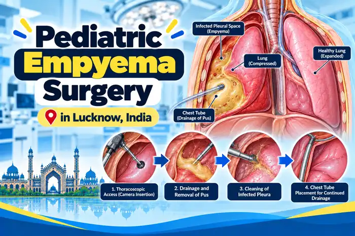
Pediatric Empyema Surgery involves procedures used to drain infected fluid or pus from the space between the lungs and the chest wall, called the pleural space. The goal is to control infection, help the affected lung expand, and improve breathing when antibiotics and other medical treatments alone are insufficient.
Depending on the stage and severity of the infection, treatment may involve chest tube drainage, drainage combined with intrapleural medicines, video-assisted thoracoscopic surgery (VATS), or open decortication. The appropriate approach depends on the child’s clinical condition, imaging findings, thickness of the infected material, and whether the lung is prevented from expanding by a thick fibrous layer.
Not every child with empyema requires an operation. Early medical treatment and drainage may be sufficient in some cases, while children with persistent collections, organized pus, or trapped lung may require more extensive surgery.
A child may require a drainage procedure or surgery when infected fluid accumulates around the lungs and interferes with breathing or does not respond adequately to initial treatment.
Surgical intervention may be considered when:
The decision is based on the child’s overall condition, clinical progress, and chest imaging.
The primary goal is to remove infected material from the pleural space and allow the affected lung to expand.
Depending on the child’s condition, treatment may aim to:
The exact procedure depends on the stage of empyema and the child’s response to antibiotics and drainage.
The procedure is performed using an approach selected according to the child’s condition and the extent of the pleural infection.
Before intervention, the team evaluates chest imaging, breathing, oxygen requirements, and other relevant clinical findings. General anesthesia is commonly used for VATS and open decortication, while chest tube placement may be performed using an appropriate anesthetic or sedation plan.
The procedure may involve:
Antibiotics remain an important part of treatment and are continued according to the child’s clinical condition and microbiological findings.
Chest tube drainage involves placing a flexible tube through the chest wall into the pleural space to remove infected fluid or pus.
The tube is connected to a drainage system, which may also help remove air when necessary. The team monitors the amount and character of the drainage and assesses whether the lung is expanding.
Chest tube drainage may be appropriate when the collection can be drained effectively without more extensive surgery.
In selected children, medicines called intrapleural fibrinolytics may be administered through the chest tube to help break down fibrin strands and improve drainage of loculated fluid.
If drainage remains inadequate, the surgical team may consider VATS or another intervention.
VATS is a minimally invasive surgical procedure used to drain infected material and clear the pleural space through small incisions in the chest.
A thoracoscope, which is a camera-equipped instrument, allows the surgeon to view the pleural cavity. Specialized instruments are used to remove pus, break down loculations, and clear infected material.
VATS may be considered when:
Potential advantages in suitable children include smaller incisions and less chest wall disruption than open surgery. However, VATS is not appropriate for every case, and the surgeon may need to convert to an open operation if safe clearance cannot be achieved.
Open decortication is a surgical procedure in which the surgeon removes a thick, restrictive fibrous layer surrounding the lung when it prevents adequate expansion.
The operation generally involves an incision in the chest to access the pleural space. The surgeon removes the organized infected material and carefully separates the restrictive layer from the lung surface where appropriate.
Open decortication may be considered when:
The objective is to release the trapped lung while protecting the lung tissue and nearby structures.
The choice depends on the extent of infection, the degree of organization, the child’s condition, and the surgeon’s assessment.
| Feature | VATS | Open Decortication |
|---|---|---|
| Access | Small chest incisions | Larger chest incision |
| Main use | Drainage, clearance of pus and loculations, selected decortication | Extensive clearance and removal of restrictive pleural peel |
| Recovery | May be shorter in suitable cases | May involve more postoperative discomfort and recovery time |
| Suitability | Depends on anatomy and disease stage | May be preferred for extensive or complex disease |
| Conversion | May be necessary if minimally invasive clearance is inadequate | Not applicable once an open approach is selected |
Neither approach is best for every child. The priority is effective infection control and safe lung expansion.
The timing depends on the child’s symptoms, the extent of the collection, and response to treatment.
Early management generally includes antibiotics and assessment of whether drainage is needed. Children with significant pleural collections, respiratory compromise, or inadequate response to initial drainage may need intervention without unnecessary delay.
More organized empyema may require VATS or decortication. The treating team determines the timing according to the child’s clinical condition and imaging findings.
Empyema drainage and surgery are established treatment options, but all procedures carry risks.
The risks depend on the severity of infection, the child’s respiratory condition, the stage of empyema, and the procedure performed.
Before surgery, the team evaluates breathing, oxygen requirements, hydration, blood tests, imaging, and other medical concerns. Antibiotics and supportive treatment are provided as required.
Children with severe infection or significant respiratory distress may need close monitoring in a pediatric intensive care setting.
Potential benefits include:
The outcome depends on the extent of infection, underlying lung condition, and effectiveness of treatment.
Possible risks include:
The surgical team explains the relevant risks and the plan for managing complications before the procedure whenever circumstances permit.
Recovery depends on the severity of infection and the procedure performed.
After surgery, the child is monitored for breathing difficulty, fever, pain, drainage, and lung expansion. A chest tube may remain in place until drainage has sufficiently reduced and the team considers removal appropriate.
Recovery may include:
Children with severe infection or extensive decortication may require a longer hospital stay.
Parents should follow the surgical team’s instructions carefully after discharge.
Important measures include:
Seek prompt medical attention if the child develops recurrent fever, worsening chest pain, increasing breathlessness, wound discharge, or unusual weakness.
Severe breathing difficulty, bluish lips, or a sudden deterioration requires emergency medical care.
Follow-up helps the team confirm that the infection is resolving and the lungs are recovering.
The doctor may assess:
Further investigations or treatment may be necessary if symptoms persist or a residual collection is suspected.
The timing depends on the severity of the infection, the surgical approach, and the child’s recovery.
Light activity can generally resume gradually as the child improves and the treating team permits. Strenuous exercise and contact sports may need to be postponed until healing is satisfactory.
Children recovering from severe pneumonia or prolonged hospitalization may need additional time to regain their usual stamina.
Many children recover well after effective treatment of empyema. The lung may gradually re-expand, and breathing and activity tolerance can improve during recovery.
Long-term outcomes depend on the severity and duration of infection, the degree of lung involvement, and whether complications occurred.
Some children may continue to experience cough, reduced exercise tolerance, or other respiratory symptoms during recovery. Persistent symptoms should be evaluated by the treating team.
The cost varies according to the child’s condition, the procedure required, and the hospital facilities.
Factors that may affect the total cost include:
Parents should request an individualized estimate from the hospital after the treatment plan has been established.
Parents should understand the extent of infection and why drainage or surgery is being recommended.
Useful questions to discuss with the treating team include:
It refers to procedures used to drain pus from the space around a child’s lungs and, when necessary, remove restrictive tissue that prevents the lung from expanding.
No. Some children improve with antibiotics and supportive treatment. Others require chest tube drainage, intrapleural medicines, VATS, or open surgery depending on the severity and response to treatment.
VATS is a minimally invasive procedure that uses a camera and specialized instruments to drain pus and clear infected material from the pleural space.
Open decortication may be considered when thick fibrous tissue traps the lung or when extensive or complex infection cannot be adequately treated with less invasive methods.
Recovery varies according to the severity of infection, procedure performed, need for chest drainage, and the child’s overall health.
Persistent or recurrent infection is possible, although effective drainage and appropriate antibiotics help reduce this risk. New fever, chest pain, or breathing difficulty should be medically assessed.
Many children recover well, but outcomes depend on the severity of infection, the condition of the lung, and any complications. Follow-up helps assess recovery.
Dr. Muni Varma offers Pediatric Empyema Surgery in Lucknow, India. With more than 10 years of experience, he uses a careful and child-focused approach to provide appropriate evaluation, surgical planning, and follow-up care. He helps parents understand treatment options for children with empyema, including drainage procedures and surgical management when required.