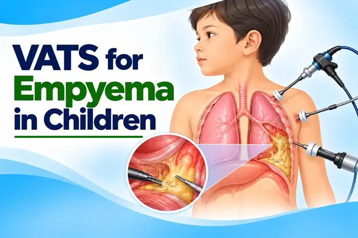
Video-Assisted Thoracoscopic Surgery (VATS) for empyema is a minimally invasive surgical procedure used to drain infected fluid and remove pus, fibrin, and infected material from the pleural space around the lungs. The surgeon uses a thin camera called a thoracoscope and specialised instruments inserted through small incisions in the chest to clean the affected area and help the lung expand.
Empyema is a condition in which pus collects in the pleural space, the area between the lungs and the inner surface of the chest wall. In children, it commonly develops as a complication of bacterial pneumonia. If infected fluid becomes thick or forms multiple pockets, antibiotics alone may not be sufficient, and drainage may be required.
VATS for empyema in children may be considered when there are complex or loculated fluid collections, persistent respiratory difficulties, or inadequate improvement with initial treatment. Depending on the child’s condition, treatment may involve antibiotics, chest tube drainage with medication to break down fibrous material, or surgery.
The choice of treatment depends on the stage of empyema, imaging findings, respiratory status, and response to previous treatment. VATS is not necessary for every child with empyema.
A pediatric surgeon may recommend VATS when infected fluid around the lungs cannot be adequately drained using less invasive methods or when the child’s condition requires surgical intervention.
VATS may be considered in children with:
The decision is made after assessing the child’s clinical condition and imaging findings. The 2026 IDSA/PIDS guidance generally suggests chest tube drainage with intrapleural fibrinolytic medication as the initial drainage approach for children with pneumonia-associated empyema who need intervention. VATS may be appropriate for selected children with extensive loculations or disease that does not respond adequately to initial treatment.
The main purpose of VATS is to remove infected material from the pleural space and help the affected lung expand.
The procedure aims to:
VATS is part of a broader treatment plan that usually includes antibiotics and supportive care. The outcome also depends on the severity of the infection and whether there is associated lung damage, such as necrotising pneumonia or a lung abscess.
VATS is performed in an operating theatre under general anaesthesia. The procedure is tailored to the amount of infected fluid, the presence of adhesions, and the condition of the lung.
The surgical team reviews the child’s symptoms, medical history, response to antibiotics, and previous drainage procedures. Imaging is used to assess the location and extent of the pleural collection.
The child receives general anaesthesia and is monitored throughout the operation. The anaesthesia team manages breathing, oxygen levels, circulation, and other vital parameters.
The surgeon makes small incisions in the chest wall. A thoracoscope with a camera is inserted through one incision, while specialised instruments are introduced through the others.
The camera provides a magnified view of the pleural space on a monitor.
The surgeon removes pus and infected fluid from around the lung. Samples may be collected for laboratory testing to help identify the bacteria responsible for the infection and guide antibiotic treatment.
If the infection has caused fibrous strands or separate pockets of fluid, the surgeon carefully separates these adhesions and removes obstructing material where appropriate. This helps clear the pleural space and improve drainage.
The surgeon assesses the lung and the cleared pleural cavity. In selected cases with a thick restrictive layer, additional procedures may be necessary to allow the lung to expand adequately.
One or more chest drains may be placed to remove residual fluid or air after surgery. The drains remain in place until the treating team determines that drainage is adequate and the lung is recovering appropriately.
After surgery, the child is monitored in a recovery area or intensive care unit, depending on the severity of the infection and respiratory condition. Antibiotics, pain relief, oxygen, and other supportive treatments are provided as needed.
The appropriate treatment depends on the stage of infection, the amount and nature of the fluid, the child’s breathing, and the response to previous treatment.
Children with a small, uncomplicated pleural effusion and no significant respiratory compromise may be treated with antibiotics and close monitoring. Drainage is considered if the infection progresses or the clinical condition warrants it.
A chest tube is inserted into the pleural space to remove infected fluid. This may be sufficient when the collection can be drained effectively.
Medication is introduced through a chest tube to help break down fibrous strands and improve drainage of loculated fluid. This is a commonly recommended initial intervention for children who need drainage for pneumonia-associated empyema.
VATS allows the surgeon to remove infected material, break down adhesions, and clear collections that cannot be managed adequately through less invasive methods. It may be used when initial drainage and fibrinolytic treatment are unsuccessful or when the anatomy and clinical condition support surgical intervention.
In selected cases, an open operation may be necessary to remove a thick restrictive layer from the lung or manage complex disease that cannot be treated adequately through VATS.
The treatment plan should be individualised. Chest tube drainage with fibrinolytic medication and VATS are both established approaches, and neither is the best option for every child.
Investigations help determine the extent of infection, identify the collection, and guide the choice of treatment.
These may include:
Not every child requires every test. The medical team selects investigations according to the child’s symptoms, clinical stability, and previous findings.
When surgery is appropriate, VATS may offer several advantages over open chest surgery.
Potential benefits include:
The benefits depend on the stage of empyema and the child’s condition. Some children require further drainage, conversion to open surgery, or additional treatment if the infection or lung damage is extensive.
VATS is an established surgical treatment for selected children with complicated empyema. However, it carries risks, particularly when the infection is severe or the lung and surrounding tissues are inflamed.
Potential complications include:
The underlying infection may also cause respiratory problems or damage to the lung. The surgical team assesses these risks and explains the expected benefits and alternatives before the procedure.
Recovery depends on the severity of infection, the child’s respiratory condition, the extent of surgery, and whether other complications are present.
The child is monitored while recovering from anaesthesia. The medical team checks breathing, oxygen levels, circulation, pain, and the amount of fluid draining from the chest.
A chest drain is often left in place temporarily. The team monitors drainage, air leakage, and lung expansion to determine when the tube can be removed.
Antibiotics are continued according to the child’s condition, culture results, and the treating team’s recommendations. The duration varies depending on the severity of infection and clinical response.
The length of hospitalisation depends on infection control, respiratory recovery, pain management, chest drain requirements, and the ability to eat and drink adequately.
Before discharge, parents receive instructions about medicines, wound care, activity, and follow-up. The treating team explains how to recognise symptoms that may indicate persistent infection or another complication.
Appropriate aftercare supports healing and helps identify complications early.
Parents should:
Parents should seek medical advice if the child develops persistent or returning fever, worsening chest pain, increasing cough, poor feeding, or new breathing difficulties. Severe breathing difficulty, bluish lips, or sudden deterioration requires emergency medical assessment.
Most children recover well after appropriate treatment for empyema, although recovery depends on the severity of the original infection and any associated lung complications.
Follow-up may include:
Not every child needs repeated imaging or specialised pulmonary testing. The follow-up plan depends on symptoms, examination findings, and the course of recovery.
A pleural effusion is an abnormal collection of fluid in the space around the lungs. It can occur with pneumonia and may be small or large.
Empyema is a more specific condition in which pus or infected fluid accumulates in the pleural space. It may develop when a pneumonia-associated effusion becomes infected and complicated.
Not every pleural effusion is empyema, and not every child with a pleural effusion needs surgery. The treatment depends on the fluid’s characteristics, the child’s breathing, and the response to antibiotics and drainage.
The cost of VATS for empyema varies according to the hospital, the child’s medical condition, the complexity of the infection, and the duration of hospitalisation.
Factors that may influence the overall cost include:
Children with severe infection or associated lung complications may need more extensive treatment. Parents should request an individual estimate after clinical evaluation so the expected costs can be explained clearly.
Before surgery, parents should understand why VATS has been recommended and whether other treatment options are appropriate.
Important points to discuss include:
Parents should share details of the child’s medicines, allergies, previous illnesses, and any treatment already received for pneumonia or pleural infection.
VATS is a minimally invasive operation that uses a camera and specialised instruments inserted through small chest incisions to drain infected fluid and remove pus or fibrous material from the space around the lungs.
No. Some children recover with antibiotics alone, while others need chest tube drainage with or without fibrinolytic medication. VATS may be recommended when drainage is inadequate, collections are extensively loculated, or the disease does not respond to initial treatment.
The best approach depends on the child’s condition. Current guidance generally supports chest tube drainage with fibrinolytic medication as the initial drainage treatment for many children with pneumonia-associated empyema. VATS is an important option for selected children with extensive loculations or inadequate response to initial treatment.
VATS is a minimally invasive operation, but it still requires general anaesthesia and specialist surgical care. The procedure may be necessary to control a complicated pleural infection and should not be considered minor simply because the incisions are small.
Recovery varies according to the severity of infection, lung condition, and the need for chest drainage or antibiotics. The hospital stay and return to normal activities are determined by the child’s clinical progress.
A chest drain is commonly used after surgery to remove residual fluid or air. It is removed when the treating team determines that drainage and lung expansion are satisfactory.
Persistent or recurrent infection is possible, although treatment aims to clear the infected collection. Follow-up is important, especially if fever, cough, chest pain, or breathing difficulties return.
Potential risks include bleeding, lung injury, persistent air leakage, residual fluid, and the need for further treatment. The individual risk depends on the severity of the infection and the child’s overall health.
Many children recover well, but outcomes depend on the severity of the infection and whether lung damage or other complications are present. The treating team monitors recovery and recommends further assessment when necessary.
Parents should contact the treating team if fever returns, chest pain worsens, or the child develops increasing cough or breathing difficulty. Severe breathing difficulty or sudden deterioration requires emergency assessment.
Dr. Muni Varma offers VATS for Empyema in Children in Lucknow, India. With more than 10 years of experience, he uses a careful and child-focused approach to provide appropriate evaluation, surgical planning, and follow-up care. He helps parents understand the available treatment options, the role of minimally invasive surgery, and the expected recovery for children requiring treatment for empyema.